![]()
Model 373PVA, biopsy training phantom
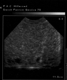
needle
in a lesion
|
|
|
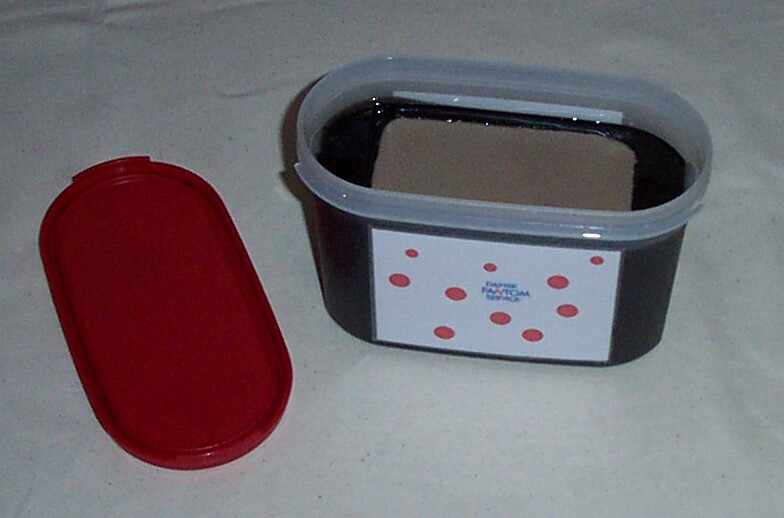
Model 373PVA, biopsy training phantom
|
|
|
||
|
|
||
|
|
||
|
|
||
|
|
||
|
|
Testing of new needles and education of young colleagues may cause unnecessary inconvenience to the patients. The biopsy phantom was developed to minimize problems of this kind.
The phantom consists of a liver tissue mimicking material containing echo poor spheres of six and ten mmØ. The material of the lesions contains red dye, so that it can be identified even in fine needle biopsies. When thin needles are used, up to thousand biopsies can be taken from this phantom. After a biopsy, air may be trapped in the biopsy canal just as in patients. But in the phantom it may take a few days to disappear.
Dimensions: 18cm * 12 cm high. Weight: 1,2 kg
Background mass: Sound speed 1540 m/sec 15 m/sec. Attenuation, backscattering and density: not specified.
Low contrast objects: Sound speed and density as background.
The phantom can be lifted out of the box and placed on the lid, so that the objects can be accessed from different directions.
Use scanning jelly. Avoid pressing the transducer into the phantom. The lifetime is dependent on the mechanical load.
Select a lesion.
Take a biopsy from the lesion.
Look at the color of the biopsy to see if the biopsy was taken from the expected lesion.